Submitted by Administrator on Wed, 12/03/2014 - 10:54
A collaborative project between the Institute of Astronomy and the CRUK Cambridge Institute.
We investigate methods for the quantitative interrogation of breast cancer histopathology using sophisticated image-processing techniques. We are developing a high-throughput image analysis platform to rapidly analyse samples from large clinical studies. This includes image processing of tissue microarray (TMA) slides as well as statistical analysis of output results.
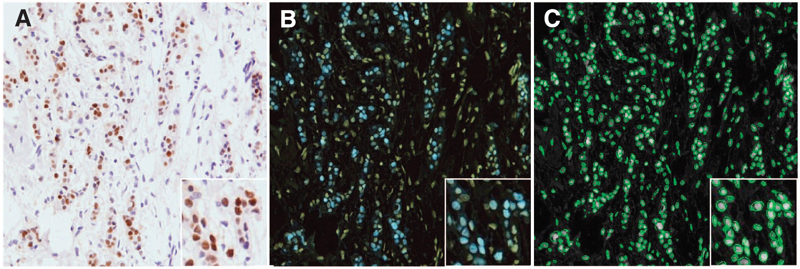
Image legend:
(A) Example image from nuclear ER staining with Allred manual score of intensity 3 and proportion 5.
(B) Converted to an astro-format and inverted.
(C) Automatic segmentation at the nuclear level with each green ellipse denoting a potential nucleus for further scoring and statistical analysis.
Image credit: Ali H. R. et al., 2013, British Journal of Cancer, 108(3):602-12.

